Challenge Open Access
Electrocardiographic Imaging of Myocardial Infarction: The PhysioNet/Computing in Cardiology Challenge 2007
Published: Jan. 2, 2007. Version: 1.0.0
Papers from the PhysioNet/CinC Challenge 2007 (Dec. 4, 2007, midnight)
Papers by the participants in the PhysioNet/Computers in Cardiology Challenge 2007 are now available, and the reference interpretations used to score the Challenge entries have also been posted.
Winners of the PhysioNet/CinC Challenge 2007 (Oct. 3, 2007, midnight)
We congratulate the winners of the PhysioNet / Computers in Cardiology Challenge 2007: Mohamed Mneimneh, Hamid SadAbadi, and Masood Ghasemi. Thanks to all who participated, and who contributed data and expertise to make this event possible.
More news
Data corrected for the PhysioNet/CinC Challenge 2007 (Sept. 21, 2007, 10:48 a.m.)
The long-axis LV model of case 3, needed for the additional challenge event 5, has been corrected. The deadline for submitting entries for event 5 only has been extended to noon GMT on Thursday, 27 September 2007. The deadline for all other events in this challenge remains noon GMT on Sunday, 23 September 2007.
Geometric models available for the PhysioNet/CinC Challenge 2007 (July 6, 2007, 10:57 a.m.)
Detailed geometric models of the heart, lungs, and torso, including a long-axis LV model, are now available for case 3 only. These have been prepared by A. van Oosterom and P. van Dam, who have also prepared a similarly detailed analysis of the infarct geometry for this case based on gadolinium-enhanced MR images. An additional challenge event that has been made possible by this analysis. The detailed models, and Matlab software for viewing them, are available here.
PhysioNet/CinC Challenge 2007 (Jan. 20, 2007, midnight)
The eighth annual PhysioNet/Computers in Cardiology Challenge has begun. This year's Challenge embraces the disciplines of both electrocardiography and (for the first time) imaging. Participants will estimate the location and extent of infarcts using ECG maps, and their estimates will be evaluated using "gold standard" measurements of gadolinium-enhanced MRI data.
Please include the standard citation for PhysioNet:
(show more options)
Goldberger, A., Amaral, L., Glass, L., Hausdorff, J., Ivanov, P. C., Mark, R., ... & Stanley, H. E. (2000). PhysioBank, PhysioToolkit, and PhysioNet: Components of a new research resource for complex physiologic signals. Circulation [Online]. 101 (23), pp. e215–e220.
Introduction
The aim of Challenge 2007 is to establish how well one can characterize the location and extent of moderate to large, relatively compact infarcts using electrocardiographic evidence (supplemented by a model of the torso geometry and conductivity), in comparison with a "gold standard" expert analysis of gadolinium-enhanced MRI data.
Background
Any aspect of clinical medicine is best served by similarly excellent methods for both diagnosis and therapy. Regarding the common clinical problem of ischemic heart disease, the diagnostic methods are currently not as well developed as the therapeutic interventions. This "mismatch" exists despite clear understanding of both the underlying basic science and the pathophysiology of ischemic heart disease and of its primary clinical manifestation, myocardial infarction. Two of the reasons for this mismatch are disconnections between basic and clinical scientists and disconnections between clinical applications of the multiple available individual diagnostic methods, for example, electrocardiography and myocardial imaging.
This year's challenge, the eighth in the series, embraces the disciplines of both electrocardiography and (for the first time) imaging. Although we expect this challenge to be difficult, we also expect that it will attract the interest of a very broad portion of the PhysioNet and CinC communities.
Challenge 2007 Development
The topic of this year's challenge, Electrocardiographic Imaging of Myocardial Infarction, was proposed by Galen Wagner (Duke University), chairman of the local organizing committee for the 2007 meeting of Computers in Cardiology (to be held in Durham, NC, USA from Sunday, 30 September through Wednesday, 3 October 2007). The VVRED (Virtual Visual Reconstructed Electrocardiographic Display) Group, led by Galen Wagner and by Rob MacLeod (University of Utah) has collected the data, and has developed the Challenge in collaboration with George Moody (MIT), representing PhysioNet.
VVRED is an informal group of investigators that has had biannual meetings hosted by Fred Kornreich (Free University of Brussels) for the purpose of facilitating advances in ECG imaging [1]. During the 2nd VVRED meeting in June 2006, the group discussed the proposed challenge topic and accepted the role of assisting in its formal development. VVRED Group member Adriaan van Oosterom (University of Lausanne) volunteered to work with Galen Wagner and the MALT (Magnetic and Electrocardiographic Technologies) Group at the Glasgow Western Infirmary. The MALT studies have acquired serial paired ECG and cardiac MRI data in patients with acute myocardial infarction. The first of these studies [2] investigated the relationship between anatomic and electrical cardiac axis; the second (submitted for publication) addressed the limits of current ECG criteria for acute MI diagnosis.
VVRED Group member Milan Horáček (Dalhousie University) provided body surface potential mapping (BSPM) capability for MALT patients returning for their one-year follow-up MRI, and he is now conducting a study aimed at delineating healed infarcts based on body surface potential maps in 40 of these patients. He has worked with Galen Wagner, MALT physicist John Foster (University of Glasgow), and Adriaan van Oosterom to present this problem as the PhysioNet/CinC Challenge 2007.
The challenge is open to a broad range of participants with and without experience in the area of electrocardiographic imaging. Participants from the clinical world (the non-modelers) and from the signal-processing world (the statistical, database-based approaches) are most welcome. The challenge was introduced during the CinC 2006 meeting in Valencia, Spain and is now being made available on the PhysioNet web site.
The Challenge Data
All of the challenge data are available in the data/ subdirectory.
The MALT sub-study patients were first inventoried to determine the distribution of infarcts in the three major locations according to Selvester ECG criteria [3]. Based on the inventory and on the availability of all necessary data, the VVRED Group selected two "training" cases and two "test" cases, in each case with a moderate to large and relatively compact infarct. Although the labor-intensive nature of the data preparation process has necessarily resulted in a small number of training and test cases, we expect that this challenge will encourage development of larger data sets to support followup studies of promising methods.
Organization of the data
All of the challenge data are available here. Reference interpretations of the training cases are available here.
The BSPM data, consisting of ECG data for 352 torso-surface sites, are provided in easy-to-read ASCII format for a single averaged PQRST complex sampled at 2kHz. These data contain the standard 12 leads, the 7 unweighted Frank leads and the Frank orthogonal XYZ leads (from which the vector loops may be constructed) as subsets.
The anatomic locations of the 120 recording electrodes, and the locations of all 352 nodes on the torso surface for which ECG data are available, are provided as well. For case 3 (one of the two test cases) only, Fady Dawoud (Dalhousie University) has provided customized torso and ventricular geometry.
To visualize the torso surface geometry and the BPSM data, you can use the map3d program, freely available from the University of Utah, SCI Institute. Commands for using map3d on the challenge data are included in the data directory.
Selected MRI sections through the heart from apex to base are also available. These images are from the patient's initial MRI study (outlines without gadolinium enhancement) to prevent disclosure of the infarct location based on shrinkage of the involved LV region during the later "remodeling" process. The data from the late enhancement image using gadolinium are provided for the training-set patients only.
Reporting Results and Scoring
- First, download this sample entry form.
- For each of the two test cases, participants must estimate one or more of the following:
- the extent of the infarct (the percentage of myocardial mass that is infarcted)
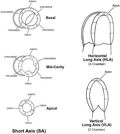
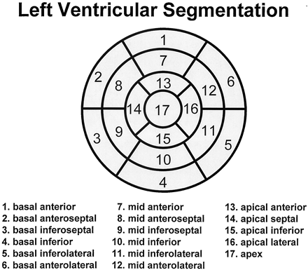
- the set of myocardial segments containing infarcted tissue
- the myocardial segment containing the centroid ("center of mass") of the infarct (using 17-segment segmentation; see figures 3 and 4 in [14], reproduced here)
- Send the filled-in entry by email to webmaster@physionet.org, with the subject line "Challenge-2007-entry". Entries will be accepted until noon GMT on 23 September 2007.
- Entries will be compared against the "gold standard" (gadolinium enhanced images). The gold standard images are provided here for the two training cases only. The gold standard images for the two test cases are available only to the challenge organizers, who will score all entries; their decisions are final.
- Scores will be based on:
- EPD: Percentage discrepancy between the extent of the infarct as estimated and as determined from the gold standard.
- SO: Overlap between the sets of infarct segments as estimated and as determined from the gold standard.
- CED: Distance between the centroid of the infarct as estimated and as determined from the gold standard.
- You will receive your scores by return email. The first scores will be sent on or about 27 April 2007; scores for entries received after that date will usually be sent within a day of receipt.
Given the estimated and gold standard sets of infarct segments, "overlap" (SO) is defined as the number of segments in both sets divided by the number of segments in either set (a value between 0 and 1, where 1 is a perfect match and 0 indicates that the sets are completely disjoint). Distance between the centroid estimates (CED) is defined as the number of segment boundaries crossed along the shortest path connecting the estimates.
You will receive up to three scores (EPD, SO, and CED) for your entry, but each score represents the sum of the subscores obtained for each of the two test cases. Individual subscores will not be made available until after the end of the Challenge in September.
Events and Awards
If you submitted an abstract describing your work on the challenge no later than 7 May 2007 to Computers in Cardiology, you may be eligible for one or more awards that will be presented during the final plenary session of the conference on Wednesday, 3 October 2007.
Three awards of US$250 each will be given to the participants who achieve the best (lowest) EPD, SO, and CED scores, and a fourth award of US$250 will be given to the participant who achieves the best average rank among all participants with respect to the other three events.
New event added 6 August 2007:
Participants are invited to determine which nodes in the long-axis LV model of case 3 lie above infarcted tissue. To enter this event, send a list of these nodes to webmaster@physionet.org, with the subject line "Challenge-2007-event-5-entry". Entries will be scored using an overlap measure similar to that defined for SO above. An award of US$250 will be provided to the most successful participant in this event.
Frequently Asked Questions
This is a hard problem, and I won't be able to get final results in time for the abstract deadline. What needs to be submitted in the abstract?
[The abstract deadline has now passed, so this question and its answer are moot.]
Briefly, you will need to submit a short (~300 words) abstract, which may not include illustrations, by 7 May.
It would be ideal if you have submitted rough results for the Challenge problem no later than 3 May, so they can be scored and so that you can report your score in your abstract. If this is not possible, please discuss in your abstract relevant studies you have performed to date, touching on any novel aspects of your methods and quoting whatever results you have. Generally abstracts containing only "we will show ..." without any results are not accepted, so it is very important that you demonstrate in the abstract that you have actually accomplished something already. Given the complexity of the challenge and the short time available for working on it, the abstract reviewers will certainly make some allowance for those who are unable to obtain results on the challenge problem by the abstract deadline, if it appears they will be able to do so before the conference in September.
You may submit entries at any time until the final deadline of noon GMT on 23 September. Each entry will be scored, and you may attempt to improve your score by submitting up to two revised entries. Only two revisions (three entries in all) are allowed in this challenge, since only a small number would be needed to obtain a good score simply by guessing, and we want to avoid that possibility!
You may choose any of your (up to three) entries as the basis for ranking, by sending an email specifying your choice on or before noon GMT on 23 September. Please remember that it may take up to 24 hours after submitting an entry to receive scores, so try to submit your last entry at least a day before the deadline if you think you may want to exercise your choice. Your last entry will be used for ranking unless you specify otherwise.
If my abstract is accepted, what else must I do?
In September, if your abstract is accepted, you will be expected to submit a four-page paper, which may be illustrated, for publication in Computers in Cardiology on-line and in print. You will also be expected to attend the conference (30 September - 3 October 2007 in Durham, North Carolina, USA) and to present your work in one of the scientific sessions of the conference, either as a poster or as a 10-minute oral presentation. Your paper and presentation should include your final results.
If you will need a visa in order to travel to the USA for the conference, please apply for it as soon as possible to avoid disappointment. The process of obtaining a visa to visit the USA has become very complicated and very slow in recent years.
Why don't you have a challenge about ...?
Each year, we receive many suggestions for challenge topics. We encourage you to contact us with further suggestions.
References
- MacLeod R, Kornreich F, van Oosterom A, Rautaharju P, Selvester R, Wagner GS, Zywietz C. Report of the first virtual visualization of the reconstructed electrocardiographic display symposium. J Electrocardiol 2005;38:385-399.
- Engblom H, Foster JE, Martin TN, Groenning BA, Pahlm O, Dargie HJ, Wagner GS, Arheden H. The relationship between electrical axis by 12 lead electrocardiogram and anatomical axis of the heart by cardiac magnetic resonance in healthy subjects. Am Heart J 2005 Sep;150:507-512.
- Anderson WD, Wagner NB, Lee KL, White RD, Yuschak J, Behar VS, Selvester RH, Ideker RE, Wagner GS. Evaluation of a QRS scoring system for estimating myocardial infarct size. VI. Identification of screening criteria for non-acute myocardial infarcts. Am J Cardiol 1988;61:729-733.
- Oostendorp TF, Nenonen J, and Korhonen P. Non-invasive estimation of the activation sequence of the heart in the presence of old myocardial infarctions: comparison to invasive patient data. J Electrocardiol 2002;35 Suppl:75-80.
- Hoekema R, Uijen GJH, and van Oosterom A. Geometrical aspects of the inter-individual variability of multilead ECG recordings, IEEE Trans Biomed Eng 2001; BME-48:551-559
- R.M. Gulrajani, P. Savard, and F.A. Roberge. The inverse problem in electrocardiography: Solutions in terms of equivalent sources. Crit. Rev. Biomed. Eng., 16:171-214, 1988.
- R.M. Gulrajani, F.A. Roberge, and P. Savard. The inverse problem of electrocardiography. In P.W. Macfarlane and T.D. Veitch Lawrie, editors, Comprehensive Electrocardiology, pages 237-288. Pergamon Press, Oxford, England, 1989.
- Y. Rudy and B.J. Messinger-Rapport. The inverse solution in electrocardiography: Solutions in terms of epicardial potentials. Crit. Rev. Biomed. Eng., 16:215-268, 1988.
- Y. Rudy and H. Oster. The electrocardiographic inverse problem. Crit. Rev. Biomed. Eng., 20:22-45, 1992.
- R.S. MacLeod, R.M. Miller, M.J. Gardner, and B.M. Horáček. Application of an electrocardiographic inverse solution to localize myocardial ischemia during percutaneous transluminal coronary angioplasty. J. Cardiovasc. Electrophysiol., 6:2-18, 1995.
- R.S. MacLeod and D.H. Brooks. Recent progress in inverse problems in electrocardiology. IEEE Eng. in Med. & Biol. Soc. Magazine, 17(1):73-83, January 1998.
- A.J. Pullan, L.K. Cheng, M.P. Nash, C.P. Bradley, and D.J. Paterson. Non-invasive electrical imaging of the heart - theory and model development. Annal. Biomed. Eng., 29(10):817-836, 2001.
- C. Ramanathan, R.N. Ghanem, P. Jia, K. Ryu, and Y. Rudy. Noninvasive electrocardiographic imaging for cardiac electrophysiology and arrhythmia. Nat Med, 10(4):422-428, April 2004.
- M.D. Cerqueira, N.J. Weissman, V. Dilsizian, A.K. Jacobs, S. Kaul, et al. Standardized myocardial segmentation and nomenclature for tomographic imaging of the heart. Circulation, 105:539-542, 2002.
- G. Wagner, G. Bub, P. Kohl, and F. Pillekamp. Electrocardiography and imaging. J Electrocardiol 40:S66-S70, 2007.
Challenge Results
The winners of the Challenge were announced at the concluding plenary session of Computers in Cardiology 2007. They are Mohamed Mneimneh (Marquette University, Milwaukee, USA), Hamid SadAbadi (K.N. Toosi University of Technology, Tehran, Iran), and Masood Ghasemi (K.N. Toosi University of Technology, Tehran, Iran). Congratulations to them, and to all of the participants who tackled this very difficult challenge. Final results for all participants show that case 4 was particularly challenging. Interestingly, the best results were obtained from the ECG signals alone, without reference to the patient-specific torso models made possible from the MR images, suggesting that further improvement should be possible using the additional information.
Papers
These papers were presented at Computers in Cardiology 2007. Please cite this publication when referencing any of these papers. These papers have been made available by their authors under the terms of the Creative Commons Attribution License 2.5 (CCAL). We wish to thank all of the authors for their contributions.
These papers were written by participants in the Challenge, who presented their work during the special challenge session at Computers in Cardiology in Durham, North Carolina, on Tuesday, 2 October 2007.
Model-Based Approach to the Localization of Infarction
D Farina, O DösselUsing Inverse Electrocardiography to Image Myocardial Infarction
FD DawoudBody Surface Potential Mapping for Detection of Myocardial Infarct Sites
P Zarychta, FE Smith, ST King, AJ Haigh, A Klinge, D Zheng, S Stevens, J Allen, A Okelarin, P Langley, A MurrayRPS/GMM Approach toward the Localization of Myocardial Infarction
MA Mneimneh, RJ PovinelliElectrocardiographic Imaging of Myocardial Infarction Using Heart Vector Analysis
M Ghasemi, A Jalali, H SadAbadi, M Atarod, H Golbayani, P Ghorbanian, A GhaffariVariation of ECG Features on Torso Plane: An Innovative Approach to Myocardial Infarction Detection
H SadAbadi, A Jalali, M Ghasemi, P Ghorbanian, M Atarod, H Golbayani, A Ghaffari
Access
Access Policy:
Anyone can access the files, as long as they conform to the terms of the specified license.
License (for files):
Open Data Commons Attribution License v1.0
Corresponding Author
Files
Total uncompressed size: 0 B.
Access the files
- Download the ZIP file (97.5 MB)
- Access the files using the Google Cloud Storage Browser here. Login with a Google account is required.
-
Access the data using the Google Cloud command line tools (please refer to the gsutil
documentation for guidance):
gsutil -m -u YOUR_PROJECT_ID cp -r gs://challenge-2007-1.0.0.physionet.org DESTINATION
-
Download the files using your terminal:
wget -r -N -c -np https://physionet.org/files/challenge-2007/1.0.0/
-
Download the files using AWS command line tools:
aws s3 sync s3://physionet-open/challenge-2007/1.0.0/ DESTINATION
| Name | Size | Modified |
|---|---|---|
| data | ||
| papers | ||
| entry.txt (download) | 210 B | 2019-04-17 |
| final-scores (download) | 1.3 KB | 2019-04-17 |
| gold-standard.txt (download) | 675 B | 2019-04-17 |
| seg1.png (download) | 24.9 KB | 2019-04-17 |
| seg2.png (download) | 50.3 KB | 2019-04-17 |
| segmentation.html (download) | 585 B | 2019-04-17 |
| training-set.txt (download) | 418 B | 2019-04-17 |