Database Open Access
STAFF III Database
Published: Jan. 31, 2017. Version: 1.0.0
New Database Added: STAFF-III Database (Jan. 31, 2017, midnight)
The STAFF-III database was acquired during 1995-96 at Charleston Area Medical Center (WV, USA) where single prolonged balloon inflation had been introduced to achieve optimal results of percutaneous transluminal coronary angiography (PTCA) procedures, replacing the typical series of brief inflations. The database consists of standard 12-lead ECG recordings from 104 patients.
Please include the standard citation for PhysioNet:
(show more options)
Goldberger, A., Amaral, L., Glass, L., Hausdorff, J., Ivanov, P. C., Mark, R., ... & Stanley, H. E. (2000). PhysioBank, PhysioToolkit, and PhysioNet: Components of a new research resource for complex physiologic signals. Circulation [Online]. 101 (23), pp. e215–e220.
Introduction
The STAFF III database was acquired during 1995–96 at Charleston Area Medical Center (WV, USA) where single prolonged balloon inflation had been introduced to achieve optimal results of percutaneous transluminal coronary angiography (PTCA) procedures, replacing the typical series of brief inflations. The lead investigator Dr. Stafford Warren designed the study protocol together with Dr. Galen Wagner at Duke University Medical Center (Durham, NC, USA); Dr. Michael Ringborn (Blekinge Hospital, Karlskrona, Sweden) was responsible for data acquisition. The database consists of ECG recordings from 104 patients, accounting for substantial inter-patient variability in reaction to prolonged balloon inflation as well as variability of heart rhythm and waveform morphology. Only patients receiving elective PTCA in one of the major coronary arteries were included. Patients suffering from ventricular tachycardia, undergoing an emergency procedure, or demonstrating signal loss during acquisition, were excluded.
Data Collection
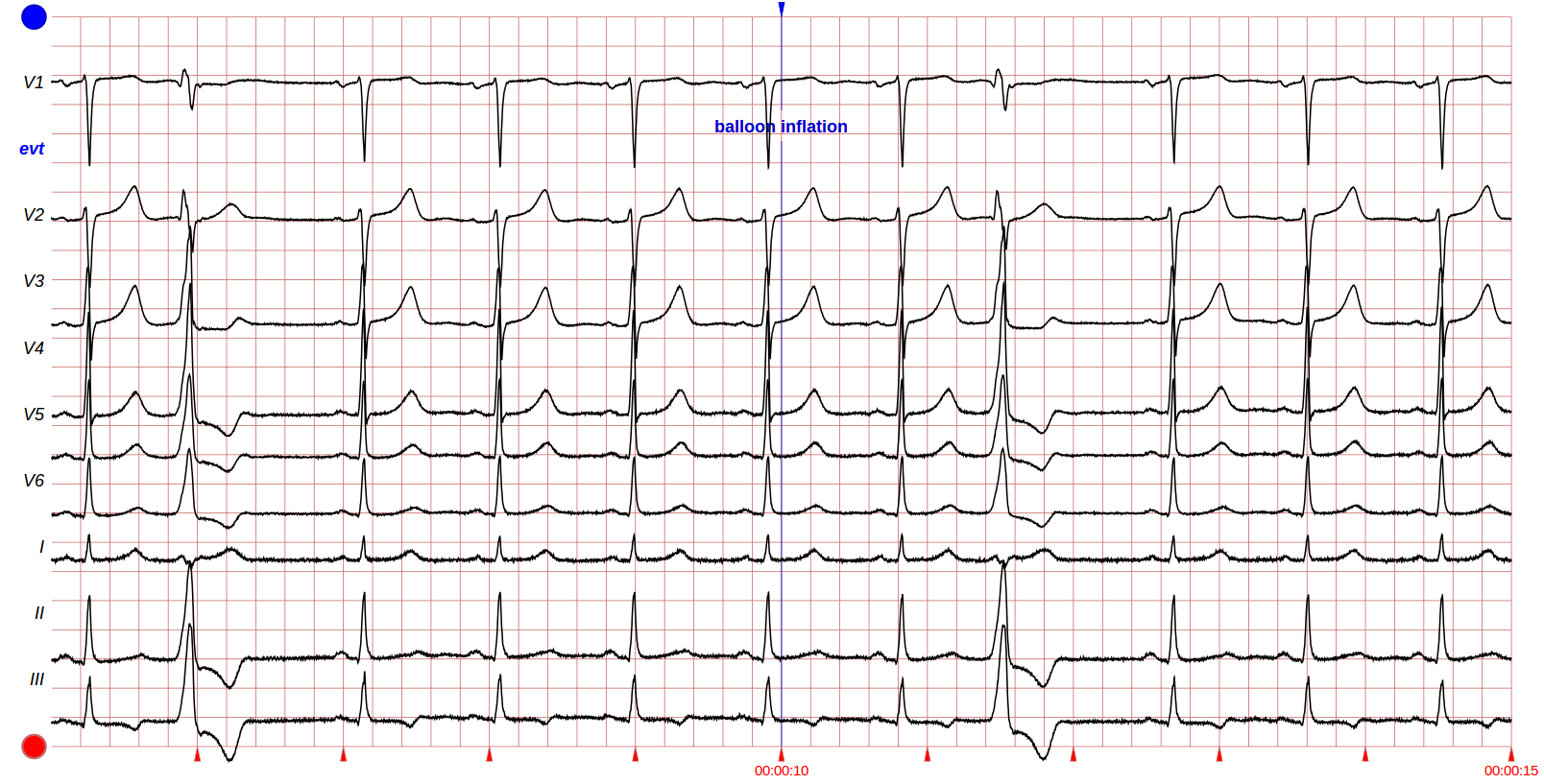
The standard procedure was defined as follows. Pre-inflation (baseline) ECGs were acquired for 5 min at rest in supine position in either a relaxing room or the catheterization laboratory, or both, prior to any catheter insertion. Inflation ECGs were acquired up to five times in each patient. The mean inflation time was 4 min 23 s, ranging from 1 min 30 s to 9 min and 54 s. In 86 inflations, the balloon did not go up immediately at the beginning of the recording, but after 4 to 205 s into the recording. Moreover, in some cases, the balloon went down before the recording ended, with a postinflation period >60 s in all but 11 inflations. All time instants related to balloon inflation/deflation were manually annotated. Post-inflation ECGs were acquired for 5 min at rest in supine position in either the catheterization laboratory or the relaxing room, or both.
The database contains a total of 152 occlusions in the major coronary arteries, distributed as 58 occlusions in left anterior descendent (LAD) artery, 59 in right coronary artery (RCA), 32 in left circumflex artery (LCX), and 3 in the left main (LM) artery. Based on ECG criteria, 35 patients had previous myocardial infarction.
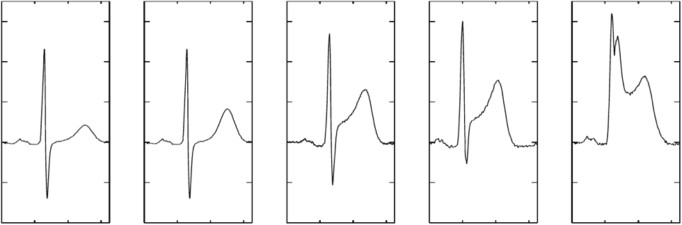
The database consists of standard 12-lead ECG data. Standard electrode placements were used for the precordial ECG leads, whereas the limb leads were obtained with the Mason–Likar electrode configuration to reduce noise originating from skeletal muscle. Data acquisition was based on custom-made equipment by Siemens–Elema AB (Solna, Sweden) with an extraordinary dynamic input amplitude range. The ECG was digitized at a sampling rate of 1000 Hz and an amplitude resolution of 0.625 µV. These specifications ensured that high-resolution digital signals could be produced which made it possible to analyze high-frequency components as well as other subtle electrophysiological phenomena.
Originally, scntigraphic images were also obtained by injecting Technetium Tc99m Sestamibi for localizing the myocardium supplied by the temporarily occluded artery. However, these images have not come to play any significant role as they are only available for a small number of all patients, and therefore not provided for download on the PhysioNet website.
Dye injections during catheterization and angiography may case changes of the ECG morphology, and the injections are therefore annotated. However, not all injections were annotated, and therefore users are advised to be cautious when ECG changes are observed that mimic the dynamics commonly observed together with annotated injections.
Data Files
All clinical information and annotations are included in the xlsx and ods format spreadsheets, including columns which describe the measurement type correspond to each recording file. The event times related to the balloon inflation/deflation and the contrast injection are also provided as WFDB annotator files.
Each recording in the database is defined by the data files *.dat and *.hea, together with an annotation file *.event which states times for balloon inflation/deflation and contrast injection (only available for certain recordings).
Comments on the original inflation annotations and the introduction of complementary information
In some files (marked in red in the spreadsheets), we identified additional inflations, not originally indicated in the original spreadsheet. This was done by checking the length of a recording vs the length of inflations; if these two numbers agreed, then we assumed that two or possibly more inflations occurred in the recording. This was the case for the following files: 7c, 11c, 39d, 53c, and 64b (2 inflations in total), and 39c (3 inflations). In addition, three recordings: 29c, 31c, and 77c, were already marked as having two inflations in the original spreadsheet. All of these extrapolated inflations are included in the .event annotation files.
In file 27c (with inflation), the time until inflation was annotated. We then assumed that the remaining part was recorded during inflation. It may be the case that the balloon went down before the end of the recording, but this information is missing.
For the second and the third inflation, if it exists, the annotated time until inflation (D0) is not strictly the time until inflation as D0 also includes the durations of previous pre-inflation(s), inflation(s), and post-inflation(s).
Pablo Laguna, Leif Sörnmo, Juan Pablo Martínez, January 19, 2017.
Contributors
Since its acquisition, the STAFF III Database has been distributed by Prof. Leif Sörnmo (Lund University, Sweden), responsible for the acquisition equipment and software. The use of the STAFF III database has broadened considerably over the years, with importance for several other research problems than high-frequency ECG analysis. Although the original study protocol of the database was designed to address a set of clinical issues, the database has turned out to be highly valuable also for developing, improving, and evaluating a wide range of signal processing techniques. This database has prompted methodological development in many areas related to ischemia, see the review by Laguna and Sörnmo (2014).
The database was prepared for PhysioNet by:
- Pablo Laguna, Prof., Zaragoza University, Spain (laguna@unizar.es)
- Leif Sörnmo, Prof., Lund University, Sweden (leif.sornmo@bme.lth.se)
- Juan Pablo Martínez, Assoc. Prof., Zaragoza University, Spain (jpmart@unizar.es)
References
The first paper listed below is the study for which the database was acquired. The other two papers review work exploring the STAFF III database, either clinical or methodological.
- Pettersson J, Carro E, Edenbrandt L, Pahlm O, Ringborn M, Sörnmo L, Warren S, Wagner G. "Spatial, individual and temporal variation of the high frequency QRS amplitudes in the 12 standard electrocardiographic leads," Am Heart J, 139(2 Pt 1):352-358, 2000.
- Warren SG, Wagner GS. "The STAFF studies of the first 5 minutes of percutaneous coronary angioplasty balloon occlusion in man". J Electrocardiol 2014;47(4):402–7.
- P. Laguna, L. Sörnmo, "The STAFF III ECG database and its significance in methodological development evaluation", J Electrocardiol, Vol. 47, pp. 408–417, 2014.
Access
Access Policy:
Anyone can access the files, as long as they conform to the terms of the specified license.
License (for files):
Open Data Commons Attribution License v1.0
Discovery
DOI (version 1.0.0):
https://doi.org/10.13026/C20P4H
Topics:
angiography
ecg
Corresponding Author
Files
Total uncompressed size: 3.2 GB.
Access the files
- Download the ZIP file (3.2 GB)
- Access the files using the Google Cloud Storage Browser here. Login with a Google account is required.
-
Access the data using the Google Cloud command line tools (please refer to the gsutil
documentation for guidance):
gsutil -m -u YOUR_PROJECT_ID cp -r gs://staffiii-1.0.0.physionet.org DESTINATION
-
Download the files using your terminal:
wget -r -N -c -np https://physionet.org/files/staffiii/1.0.0/
-
Download the files using AWS command line tools:
aws s3 sync s3://physionet-open/staffiii/1.0.0/ DESTINATION
| Name | Size | Modified |
|---|---|---|
| data | ||
| ANNOTATORS (download) | 74 B | 2017-01-26 |
| RECORDS (download) | 5.1 KB | 2017-01-26 |
| SHA256SUMS.txt (download) | 92.1 KB | 2019-02-20 |
| STAFF-III-Database-Annotations.ods (download) | 28.9 KB | 2017-01-26 |
| STAFF-III-Database-Annotations.xlsx (download) | 29.3 KB | 2017-01-26 |
| STAFF-Studies-bibliography-2016.pdf (download) | 145.5 KB | 2017-01-31 |
| STAFFIII-illustration.png (download) | 35.8 KB | 2017-01-26 |
| Signals.png (download) | 292.8 KB | 2017-01-26 |